An echocardiogram test, often called an echo or 2D echo, uses sound waves to create moving pictures of your heart. It shows the size of the heart, how well it pumps and how the valves work, without any radiation or injection in most cases. Doctors order an echocardiogram test for breathlessness, chest pain, a heart murmur or known valve disease. This guide explains the types, preparation, what happens during the scan and how to understand the report.
Key points at a glance
- An echo is an ultrasound of the heart and uses no X-rays.
- The standard test is painless and usually takes less than an hour.
- Different types of echo are used for different questions.
- A standard echo does not directly show blockages in the heart arteries.
What Is an Echocardiogram Test?
According to the Mayo Clinic, an echocardiogram is an ultrasound test that creates pictures of the heart and shows blood flow through the heart and its valves. A small probe, called a transducer, sends out sound waves, and a computer converts the returning echoes into live images. "2D echo" simply refers to the two-dimensional pictures that most echo machines produce, and some machines can also show 3D images.
Why Is an Echocardiogram Done?
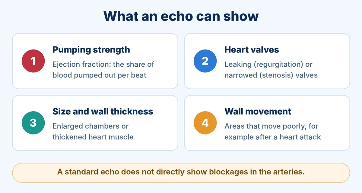
An echo shows structure and function, but a standard echo does not directly show artery blockages.
- Breathlessness or chest pain: to look for a heart cause.
- Heart failure: to see how well the heart pumps and fills. Read our guide on congestive heart failure.
- Heart murmur: to find out whether a valve problem is behind it.
- Valve disease: to check for leaking or narrowed valves, as in aortic stenosis and mitral regurgitation.
- After a heart attack: to look for areas of heart muscle that are not moving well. See heart attack symptoms, causes and treatment.
- Irregular rhythm: to look at the heart structure in conditions such as atrial fibrillation.
- Heart defects present from birth.
Types of Echocardiogram
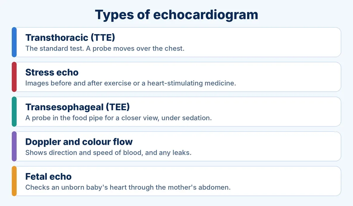
Most people have the standard transthoracic echo, where the probe stays outside the chest.
- Transthoracic echo (TTE): the standard, non-invasive test, in which the probe is moved over the chest. In some people, a contrast agent is given through an IV line to make the pictures clearer.
- Stress echo: pictures are taken before and after exercise on a treadmill or bicycle, or after a medicine that makes the heart work harder if you cannot exercise. It helps to see how the heart behaves under stress.
- Transesophageal echo (TEE): a thin probe is passed through the mouth into the food pipe, which sits right behind the heart, to get a closer view of the heart and aorta. The throat is numbed and sedation is usually given.
- Doppler and colour flow: these show the speed and direction of blood flow and highlight leaks across the valves.
- Fetal echo: checks the heart of an unborn baby through the mother's abdomen, commonly around 18 to 22 weeks of pregnancy.
How to Prepare for an Echocardiogram
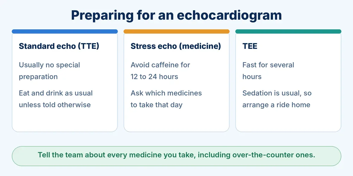
Preparation depends on the type of echo, so follow your hospital's instructions.
- Standard echo: usually no special preparation, and you can generally eat and drink as usual unless told otherwise.
- TEE: usually needs fasting for several hours. Because of sedation, arrange a ride home and plan to rest for the day.
- Stress echo with medicine: you may be asked to avoid caffeine, such as coffee, tea, chocolate and energy drinks, for 12 to 24 hours.
- All types: ask whether to take your regular medicines that day, and tell the team about every medicine you take.
What Happens During the Test?
For a standard echo you change into a gown and lie on your side or back. Small sticky patches for an ECG are often placed on your chest. The technician applies gel and presses the probe against your chest, moving it to get different views. You may be asked to hold your breath or turn on your left side for a short while. You may feel light pressure, but the test is usually painless. Most echocardiograms take less than an hour, and you can usually return to normal activities afterwards.
Are there any risks?
The ultrasound itself is considered safe. Contrast agents can rarely cause a reaction such as headache, backache or rash. A TEE may cause a sore throat or hoarseness, and rarely more serious problems, so it is done only when the closer view is needed. A medicine-based stress echo can briefly cause a fast heartbeat, flushing or a drop in blood pressure.
Understanding Your Echo Report
A cardiologist or a doctor trained in echocardiography reviews the images and writes the report, which is often ready by the next working day. It may describe:
- Ejection fraction (EF): the percentage of blood the left ventricle pumps out with each beat. A low EF means the heart is pumping weakly.
- Chamber size and wall thickness: whether any chamber is enlarged or the muscle is thickened.
- Wall motion: whether any area moves poorly, which can point to earlier damage.
- Valves: whether any valve leaks or is narrowed, and how severe it is.
Do not judge your report from a single number. The meaning of each finding depends on your symptoms and other tests, so discuss the report with your cardiologist.
What an echo cannot show
A standard echo does not directly show blockages in the heart arteries. A stress echo can give clues, but if a blockage is suspected, your doctor may advise a coronary angiogram or CT angiography. Read about coronary angiography and about signs of a heart blockage you should not ignore.
When Should You See a Cardiologist?
Consult a cardiologist if you have breathlessness, chest discomfort, swelling of the feet, palpitations or fainting, or if a doctor has heard a murmur. Chest pain with sweating or sudden severe breathlessness is an emergency. Also see the signs of an unhealthy heart.
Echocardiography and Heart Care in Jaipur
Dr. Prashant Dwivedi is an interventional and structural heart cardiologist practising at the Eternal Heart Care Centre and Research, Jaipur. Echocardiography is a key part of evaluating valve disease and heart function, and the results guide the next steps in your care. You can book an appointment for a personal assessment.
Frequently Asked Questions
Is an echocardiogram painful?
A standard echo is usually painless. You may feel light pressure from the probe. A TEE is done with throat numbing and sedation, so it is more involved.
How long does an echo take?
Most echocardiograms take less than an hour. A TEE takes longer overall because of preparation and recovery from sedation.
Do I need to fast for a 2D echo?
Usually not for a standard echo. Fasting is generally needed for a TEE. Follow the instructions given by your hospital.
Is an echocardiogram safe?
Yes, the ultrasound uses sound waves and no radiation, so it is considered safe. Small risks are linked to contrast, TEE or medicine-based stress tests, and your doctor will discuss them if they apply.
Can an echo detect a heart blockage?
A standard echo does not directly show blockages in the arteries. It can show muscle that is not moving well, and a stress echo can help. Confirming a blockage may need an angiogram or CT scan.
This article is for general educational purposes only and does not replace personal medical advice, diagnosis or treatment. Please consult a qualified cardiologist about your own condition. In an emergency, go to the nearest hospital.
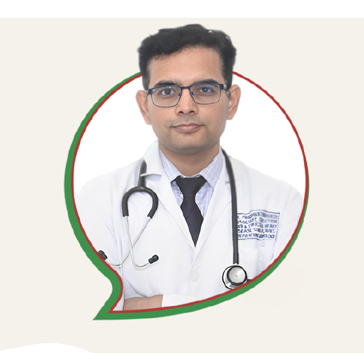
Dr. Prashant Dwivedi
Interventional and Structural Heart Cardiologist with expertise in TAVR, TMVR, MitraClip and advanced cardiac procedures.